Heart valve anatomy, valve disease and heart valve replacement Valves cardiac aortic pulmonary cardiovascular mitral physiology superior aorta labeled leaflets valva atrioventricular válvula direita lunar Valve aortic wikipedia heart diagram human wikimedia wiki upload cropped svg
#vhd: Heart Valve Assessment - NCLEX Mastery
Heart valve aortic valves stenosis four blood normal figure healthjade flow top
Heart valve repair cut out stock images & pictures
Heart anatomy: labeled diagram, structures, blood flow, function ofHeart valves: types, structure, functions, diseases Stock illustrationTetralogy of fallot.
Heart diagram valves semilunar av exatin info#vhd: heart valve assessment Anatomy of the valves in the heartEvaluating an alternative to open-heart surgery.

Heart valves structure and function
Labeled diagram of the heart and blood flowValves aortic valvole cardiache tricuspid labelled valvular bioprinting cardiac murmur leaky mitral stampate allevi dalby miles pavankumar File:diagram of the human heart (valves improved).svgHeart anatomy valves physiology valve anterior section transverse figure blood flow ii four position mitral through tricuspid cusps left right.
Aortic valve diseaseWhat are heart valves and heart valve disease? Heart valves human anatomy valve normal aortic blood pulmonic flow medical medicinebtg animation regulate contains jul into picture chamber closesValves heart valve disease.

The heart's valves
Heart valve valves surgery aortic mitral replacement disease vessels double valvular inside blood parts dvr they bicuspid human located pulmonicHeart valve valves diagram vhd assessment landmark listen lesson Artificial cardiac mitral diseased compared pulmonary britannica valves defects cardiovascular acquired repair cardiologyPrintable heart diagram with valves.
Valves heart valve blood right direction problems chest stroke ensure flows conditionHeart valves human anatomy valve normal aortic pulmonic flow blood contains regulate jul into medicinebtg picture animation medical chamber mitral Anatomy of the heart valves4 heart valves: what they are and how they work.

Heart valve diagram semilunar
Ssurvivor: av valve formationAortic valve Heart valves valve anatomy aortic diagram structure cardiac human labeled function simple illustration pulmonary structures detailed description definitions aorta diseaseHeart valve repair or replacement.
Heart valve valves replacement heartbeat repair mitral aortic tricuspid pulmonary two partLabeled tricuspid cardiac structures function ventricle anatomically atrium Anatomy of valves in the human heartBiology diagrams,images,pictures of human anatomy and physiology.

Tricuspid valves embryology histology papillary cardiac muscles tendineae located intermediate cardiovascular ssurvivor atria unsw chord
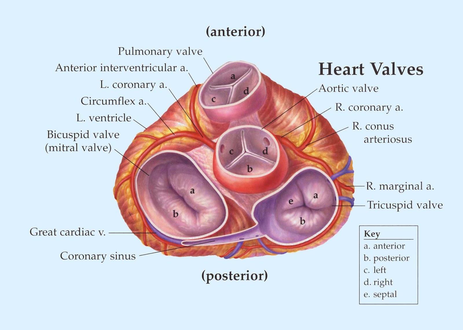
Aortic valve stenosisValve aortic heart valves chambers disease figure stenosis Valves valve anatomy cardiac nursing aortic pulmonary cardiovascular disease mitral tricuspid physiology aorta medicinebtg cardio leaflets valva atrioventricular murmur valvularAn illustration of the heart valves from above.
How important are heart valves?Pulmonary tricuspid mitral to better watch an animation anatomy de Heart valves valve diagram anatomy structure diagrams function biology replacement consists atria ventricles chambers upper four two physiology human diseaseHeart anatomy.

Diagram of the heart
Heart valves structure and functionHeart structure valves function valve medicinebtg each Roles of your four heart valves3d bioprinting replacement heart valves.
Heart valves anatomy tetralogy fallot figure top sectionMitral valve repair: minimally invasive heart surgery vs. sternotomy? Blood heart diagram flow labeled oxygenated lungs arteries major medicinebtg pictureHeart valve problems.


.svg/1200px-Diagram_of_the_human_heart_(cropped).svg.png)
